ملف:Free-living amebic infections.png
الصوره الاصليه (1,365 × 1,170 بكسل حجم الفايل: 715 كيلوبايت، نوع MIME: image/png)

الملف دا من ويكيميديا كومنز و ممكن تستعمله المشاريع التانيه. الوصف بتاعه صفحة وصف الملف هناك معروض تحت..

هذه biology الصورة / الصورتان باستعمال رسومات متجهية ملفات رسوميات شعاعية. It is recommended to name the SVG file "Free-living amebic infections.svg" - then the template Vector version available (or Vva) does not need the new image name parameter.

الخلاصة

وصف
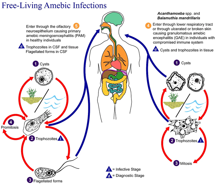
English: This is an illustration of the life cycle of the parasitic agents responsible for causing “free-living” amebic infections.

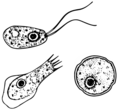
For a complete description of the life cycle of these parasites, select the link below the image or paste the following address in your address bar: http://www.dpd.cdc.gov/dpdx/HTML/FreeLivingAmebic.htm Free-living amebae belonging to the genera Acanthamoeba, Balamuthia, and Naegleria are important causes of disease in humans and animals. Naegleria fowleri produces an acute, and usually lethal, central nervous system (CNS) disease called primary amebic meningoencephalitis (PAM). N. fowleri has three stages, cysts (1) , trophozoites (2) , and flagellated forms (3) , in its life cycle. The trophozoites replicate by promitosis (nuclear membrane remains intact) (4) . Naegleria fowleri is found in fresh water, soil, thermal discharges of power plants, heated swimming pools, hydrotherapy and medicinal pools, aquariums, and sewage. Trophozoites can turn into temporary flagellated forms which usually revert back to the trophozoite stage. Trophozoites infect humans or animals by entering the olfactory neuroepithelium (5) and reaching the brain. N. fowleri trophozoites are found in cerebrospinal fluid (CSF) and tissue, while flagellated forms are found in CSF.

Acanthamoeba spp. and Balamuthia mandrillaris are opportunistic free-living amebae capable of causing granulomatous amebic encephalitis (GAE) in individuals with compromised immune systems. Acanthamoeba spp. have been found in soil; fresh, brackish, and sea water; sewage; swimming pools; contact lens equipment; medicinal pools; dental treatment units; dialysis machines; heating, ventilating, and air conditioning systems; mammalian cell cultures; vegetables; human nostrils and throats; and human and animal brain, skin, and lung tissues. B. mandrillaris however, has not been isolated from the environment but has been isolated from autopsy specimens of infected humans and animals. Unlike N. fowleri, Acanthamoeba and Balamuthia have only two stages, cysts (1) and trophozoites (2) , in their life cycle. No flagellated stage exists as part of the life cycle. The trophozoites replicate by mitosis (nuclear membrane does not remain intact) (3) . The trophozoites are the infective forms and are believed to gain entry into the body through the lower respiratory tract, ulcerated or broken skin and invade the central nervous system by hematogenous dissemination (4). Acanthamoeba spp. and Balamuthia mandrillaris cysts and trophozoites are found in tissue.
تاريخ
مصدر

This media comes from the Centers for Disease Control and Prevention's Public Health Image Library (PHIL), with identification number #3412.

Note: Not all PHIL images are public domain; be sure to check copyright status and credit authors and content providers.


العربية | Deutsch | English | македонски | slovenščina | +/−

مؤلف
  • Photo Credit:
  • Content Providers(s): CDC/Alexander J. da Silva, PhD/Melanie Moser
سماح
(إعادة استخدام الملف ده)
English: None - This image is in the public domain and thus free of any copyright restrictions. As a matter of courtesy we request that the content provider be credited and notified in any public or private usage of this image.
إصدارات أخرى

الترخيص

Public domain
هذه الصُّورة مِن إِنتاج مراكز السيطرة على الأمراض والوقاية منها (CDC)، التَّابِعة لوزارة الصِّحة والخدمات البشريَّة الأمريكيَّة، التقطت أو أُنشئت بصفتها جزءاً مِن عمل المُوظَفين الرَّسميين فيها. ولأَنَّها عملٌ من إِنتاج الحكومة الاتحادية للولايات المُتحدة الأمريكيَّة، فإِنَّ هذه الصُّورة في النِّطاق العامِّ.

سجلُّ الرَّفع الأصيل

Transferred from en.wikipedia to Commons by Optigan13.

The original description page was here. All following user names refer to en.wikipedia.
  • 2006-05-04 01:09 Keenan Pepper 518×435×8 (31658 bytes) Free-living_amebic_infections.gif, converted to [[PNG]] by [[netpbm]].

الشروحات

أضف شرحاً من سطر واحد لما يُمثِّله هذا الملف

العناصر المصورة في هذا الملف

يُصوِّر العربية

نوع الوسائط العربية

image/png

تاريخ الفايل

اضغط على الساعه/التاريخ علشان تشوف الفايل زى ما كان فى الوقت ده.

الساعه / التاريخصورة صغيرةابعاديوزرتعليق
دلوقتي09:24، 2 فبراير 2023تصغير للنسخة بتاريخ 09:24، 2 فبراير 20231,365 × 1,170 (715 كيلوبايت)Materialscientisthttps://answersingenesis.org/biology/microbiology/the-genesis-of-brain-eating-amoeba/
06:30، 20 يوليه 2008تصغير للنسخة بتاريخ 06:30، 20 يوليه 2008518 × 435 (31 كيلوبايت)Optigan13{{Information |Description={{en|This is an illustration of the life cycle of the parasitic agents responsible for causing “free-living” amebic infections. For a complete description of the life cycle of these parasites, select the link below the image

مافيش صفحات بتوصل للفايل ده.

استخدام الملف العام

الويكيات التانيه دى بتستخدم الفايل ده:

بيانات ميتا